Agilent / Varian 31-cm Horizontal Bore micro-MRI


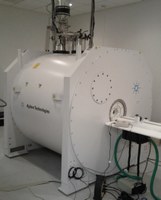
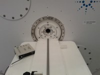
Agilent Technologies 9.4 Tesla/ 31-cm bore-hole small animal MR is a high field strength superconducting research imaging magnet, which is capable of imaging live subjects as small as mice and as large as a rabbit. This scanner produces superb 2D and 3D soft-tissue contrast due to Agilent Technologies high performance magnet technology and its operation frequency of 400 MHz. MR image quality is further enhanced by Agilent Technology’s patented active shielding technology with a drift rate less than 0.05 ppm/hr. Magnetic field homogeneity is optimized by Agilent Technologies highly stabled cryo-shimming technology. Another unique feature of this 9.4T scanner is its zero-boiling off design where the dissipating of liquid Helium is minimized, thereby reducing operational cost due to less Helium liquid refilling.
With the scanner versatile hardware design (2 transmitting and 8 receiver channels) powered by the company DirectDrive technology which enhances accurate signal digitization and timing, the scanner is a robust platform with small footprint (22,000 lb). Moreover, due to its efficient shimming technology and dynamic signal sampling provided by the DirectDrive system, it allows imaging of intricate small-scale structure with few or little aliasing artifact. Our current system includes a complete set of 3 heavy duty and efficient gradient coils (305/210 HD, 300A, 300mT/m; 205/120 HD, 300A, 600mT/m; and 115/60 HD, 200A, 1000mT/m). To further improve image signal-to-noise ratios, different designated imaging RF coil are available. Two volume Birdcage quadrature coils are used to image whole body and organ specific volume (200/254 and 72/119 mm). Five sets of phase array coils are available for acquisition of area of interest such as rodent hearts and the neurological system. A set of six surface coils is available for image acquisition of xenografted tumor models and magnetic resonance spectroscopy (MRS) data for in vivo biochemical analysis. Two specialized RF coils are also available for homogenous S/N imaging (Millipede) and utrashort time imaging (SWIFT non-echo technique).
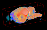
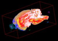
The combination of the scanner versatile design, efficient shimming, robust gradients and imaging RF coils, enhanced visualization of soft-tissue contrast in rodent pre-clinical models becomes a reality. MR modality provides investigators with a great opportunity to study in vivo disease models non-invasively. Moreover, with its capability to acquire superb soft-tissue anatomical maps, MRI/MRS is an indispensable image modality for accurate co-registration with other modalities such as PET, SPECT and optical for accurate functional and physiological mapping.