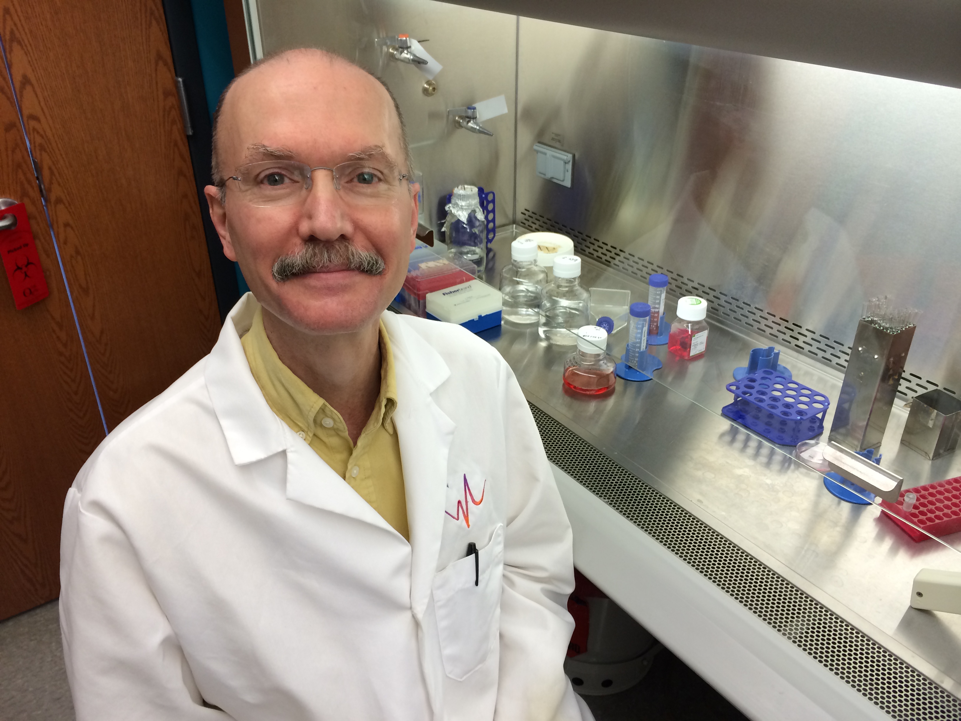
Nolan L. Boyd, Ph.D.

Nolan L. Boyd, Ph.D.
Associate Professor
Boyd Lab, Cardiovascular Innovation Institute
502.852.3169 (O)
EDUCATION
| B.S. | Mechanical Engineering | University of Texas at Arlington |
| M.S. | Bioengineering | Texas A&M University |
| Ph.D. | Bioengineering | Georgia Institute of Technology |
| Postdoc | Embryonic Stem Cell Biology | University of Georgia |
RESEARCH INTEREST STATEMENT
My lab is interested in the design and fabrication of functional tissue mimics. Tissue mimics could be useful for investigating basic biological processes, drug toxicity testing or regenerative medicine applications. The design of a mimic requires three basic components, 1) cells, 2) matrix and 3) architectural assembly.
Cells
Most organs are composed of three classes of cells, 1) the parenchyma or the cells that perform the organ function, 2) vasculature and 3) stromal cells or the tissue support cells (e.g. fibroblasts). To generate parenchymal cells we utilize pluripotent stem cells (PSC) that can differentiate into all cells of the body. PSC can be generated to be patient specific and can model genetic or metabolic pathologies. PSC have been differentiated into most of the body’s cells including cardiomyocytes, endothelium, hepatocytes, islets and neurons, to name a few. Though individual cell lineages can be derived using PSC, including endothelium, vascular mural and various stromal cells, we utilize an inherent stroma-vascular cell source possessed by everyone, adipose (i.e. fat) tissue. When adipose tissue is digested to its component cells and the adipocytes removed, what remains is referred to as the stromal vascular fraction (SVF). We have demonstrated that this heterogeneous mix of cells contains all the required components to self-assemble into a functional vasculature that interacts with implanted parenchymal cells and interfaces with the host circulation (Nunes, et al, Scientific Reports, 2013).
Matrix
Organ component cells live within a three dimensional (3D) space. 3D configurations require structural support that within the organ is provided by the extracellular matrix (ECM). The tissue mimic can utilize endogenous matrix proteins (e.g. collagens), naturally occurring matrix (e.g. alginate) or synthetic polymers (e.g. polyglycolic acid). The matrix composition and configuration can be tuned to provide specific mechanical or degradation properties.
Architectural Assembly
To generate the tissue mimic an important question is how does architectural arrangement affect function? How do developing organs self-assemble into specific arrangements? For medical applications, does a mimic have to completely recapitulate the endogenous tissue architecture to provide therapeutic function? To investigate these questions we collaborate with Dr. Stuart Williams and utilize bioprinting technology (Smith, et al, Tissue Engineering, 2004, 2007; Williams, et al, Bioprocessing, 2013) to arrange matrix and cells in complex 3D configurations.
PUBLICATIONS
- M Ramakrishnan, NL Boyd*, The adipose stromal vascular fraction as a complex cellular source for tissue engineering applications, Tissue Engineering Part B, doi: 10.1089/ten.TEB.2017.0061, PMID: 28316259 *Corresponding Author
- L Omer, EA Hudson*, S Zheng*, JB Hoying, Y Shan and NL Boyd, CRISPR correction of a homozygous LDLR mutation in familial hypercholesterolemia induced pluripotent stem cells, Hepatology Communications 1(9):886, 2017, PMID: 29130076, PMCID: PMC5677509, DOI: 10.1003/hep4.1110*Equal Contribution
LP Slomnicki, DH Chung, A Parker, T Hermann, NL Boyd & M Hetman, ribosomal stress and Tp53-mediated neuronal apoptosis in response to capsid protein of the Zika virus, Scientific Reports, 7(1):16652, 2017, PMID: 29192272, PMCID: PMC5709411, DOI: 10.1038/s41598-017-16952-8
V.M. Ramakrishnan, K.T. Tien, T.R. McKinley, B.R. Bocard, T.M. McCurry, S.K. Williams, J.B. Hoying and N.L. Boyd, Wnt5a regulates the assembly of human adipose stromal vascular fraction-derived microvasculatures, PLoS One, 11(3):e0151402, 2016. PMID: 26963616.
- V.M. Ramakrishnan*, J.Y. Yang*, B.R. Bocard, K.T. Tien, J.G. Maijub, P.O. Burchell, S.K. Williams, M.E. Morris, J.B. Hoying, R. Wade-Martins, F.D. West and N.L. Boyd, Extrachromosomal vector restores I function in genetic deficient I-derived cells, Scientific Reports, 5:13231, 2015, DOI: 10.1038/srep13231. PMID: 26307169 *Equal Contribution
- J.G. Maijub*, N.L. Boyd*, J.R. Dale, J.B. Hoying, M.E. Morris and S.K. Williams, Concentration dependent vascularization of adipose stromal vascular fraction cells. Cell Transplantation, Nov. 13, 2014. PMID:25397993 *Equal Contribution
- L.T. Kuhn, Y. Liu, N.L. Boyd, J.E. Dennis, J. Xi, X. Xin, L. Wang, H. Aguila, A.J. Goldberg, D. Rowe and A. Lichtler, Developmental engineering of bone using human embryonic stem cell-derived mesenchymal cells, Tissue Engineering, Part A, 20(1-2):365-377, 2014.
- S.S. Nunes, J.G. Maijub, L. Krishnan, V.M. Ramakrishnan, L.R. Clayton, S.K. Williams, J.B. Hoying* and N.L. Boyd*, Generation of a functional liver tissue mimic using adipose stromal vascular fraction cell-derived vasculatures, Nature Scientific Reports, 3:2141-2148, 2013. *Equal contribution
- X. Guo, S.L. Stice, N.L. Boyd and S.Y. Chen, A novel in vitro model system for smooth muscle differentiation from human embryonic stem cell-derived mesenchymal cells, American Journal of Physiology, 304 (4):C289-C298, 2013.
- N.L. Boyd*, S.S. Nunes, L. Krishnan, J.D. Jokinen, V.M. Ramakrishnan, A.R. Bugg and J.B. Hoying, Dissecting the role of human embryonic stem cell derived mesenchymal cells in HUVEC network stabilization in 3D environments, Tissue Engineering, Part A, 19 (1-2):211-223, 2013. *Corresponding Author
- M.L. Shirley, A. Venable, R.R. Rao, N.L. Boyd, S.L. Stice, D. Puett and P. Narayan, Bone Morphogenetic Protein-4 affects both trophoblast and non-trophoblast lineage-associated gene expression in human embryonic stem cells, Stem Cell Discovery, 2(4): 163-175, 2012.
- D.L. Simpson, N.L. Boyd, S.L. Stice, S.C. Dudley, Use of Human Embryonic Stem Cell Derived-Mesenchymal Cells for Cardiac Repair, Biotechnology and Bioengineering, 109 (1): 274-283, 2012.
- Boyd, N.L.*, S.S. Nunes, J.D. Jokinen, L. Krishnan, Y. Chen, K. Smith, S.L. Stice, J.B. Hoying, Microvascular Mural Cell Functionality of Human Embryonic Stem Cell Derived Mesenchymal Cells, Tissue Engineering, Part A. 17(11-12): 1537-1548, 2011. *Corresponding Author
- Dhara, S.K, Gerwe, B.A., Majumder, A., Dodla, M.C., Boyd, N.L., Machacek, D.W., Hasneen, K., and Stice, S.L.: Genetic manipulation of neural progenitors derived from human embryonic stem cells. Tissue Engineering, Part A. 15 (11): 3621-3634, 2009.
- Boyd, N.L., Dhara, S.K., Robbins, K.R. and Stice, S.L.: Human Embryonic Stem Cell Monolayer Culture for the Derivation of Fibroblastic Mesenchymal Progenitor Cells. Tissue Engineering, Part A. 15 (8): 1897-1907, 2009.
- West, F.D., Machacek, D.W., Boyd, N.L., Pandiyan, K., Robbins, K.R. and Stice, S.L.: Enrichment and Differentiation of Human Germ-like Cells Mediated by Feeder Cells and Basic Fibroblast Growth Factor Signaling. Stem Cells. 26 (11): 2768-2776, 2008.
- Dhara, S.K., Hasneen, K., Machacek, D.W., Boyd, N.L., Rao, R.R., and Stice, S.L.: Human neural progenitor cells derived from embryonic stem cells in feeder-free cultures. Differentiation. 76 (5): 454-464, 2008.
- Boyd, N.L., Dhara, S.K., Gerwe, B.A., Rao, R.R., Godbey, E.A. and Stice, S.L.: BMP4 Promotes Formation of Primitive Vascular Networks in Human Embryonic Stem Cell Derived Embryoid Bodies. Experimental Biology and Medicine. 232: 833-843, 2007.
- Stice, S.L., Boyd, N.L., Dhara, S.K., Gerwe, B.A., Machacek, D.W. and Shin, S.: Human embryonic stem cells: challenges and opportunities. Reproduction, Fertility and Development. 18 (8): 839-846, 2006.
- Pardo, S.J., Patel, M.J., Sykes, M.C., Platt, M.O., Boyd, N.L., Sorescu, G.P., Xu, M., van Loon, J.J.W. A., Wang, M.D. and Jo, H.: Simulated Microgravity Using the Random Positioning Machine Inhibits Differentiation and Alters Gene Expression Profiles of 2T3 Pre-osteoblasts. American J. Physiology-Cell Physiology. 288 (6): C1211-1221, 2005.
- Sorescu, G.P., Song, H., Tressel, S.L., Hwang, J., Dikalov, S., Smith, D.A., Boyd, N.L., Platt, M.O., Lassègue, B., Griendling, K.K. and Jo, H.: Bone morphogenic protein 4 produced in endothelial cells by oscillatory shear stress induces monocyte adhesion by stimulating reactive oxygen species production from a nox1-based NADPH oxidase. Circulation Research. 95 (8): 773-779, Oct. 15 2004.
- Boyd, N.L., Park, H., Sun, W.P., Coleman, S., Cherukuri, R., and Jo, H.: Bovine Caveolin-2 Cloning and Effects of Shear Stress on Its Localization in Bovine Aortic Endothelial Cells. Endothelium. 11 (3-4): 189-198, May-Aug. 2004.
- Boyd, N.L., Park, H., Yi, H., Boo, Y.C., Sorescu, G.P., Sykes, M. and Jo, H.: Chronic Shear Induces Caveolae Formation and Alters ERK and Akt Responses in Endothelial Cells. American J. Physiology - Heart and Circulatory Physiology. 285 (3): H1113-22, September, 2003.
- Sorescu, G.P., Sykes, M., Weiss, D., Platt, M.O., Saha, A., Hwang, J., Boyd, N., Boo, Y.C., Vega, J.D., Taylor, W.R. and Jo, H.: Bone morphogenic protein 4-produced in endothelial cells by oscillatory shear stress stimulates an inflammatory response. J. Biological Chemistry. 278 (33): 31128-35, August 15, 2003.
- Boo, Y.C., Sorescu, G., Boyd, N., Shiojima, I., Walsh, K., Du, J., and Jo, H.: Shear stress stimulates phosphorylation of eNOS at Ser1179 by Akt-independent mechanisms - Role of Protein Kinase A. J. Biological Chemistry. 277 (5): 3388-96, February 1, 2002.
- Moritomo, H., Viegas, S.F., Elder, K.W., Nakamura, K., Dasilva, M.F., Boyd, N.L. and Patterson, R.M.: Scaphoid Nonunions: a 3-Dimensional Analysis of Patterns of Deformity, J. Hand Surgery. 25 A (3): 520-8, May, 2000.
- Viegas, S.F., Yamaguchi, S., Boyd, N.L., and Patterson, R.M.: The Dorsal Ligaments of the Wrist: Anatomy, Mechanical Properties, and Function. J. Hand Surgery. 24 A (3): 456-458, May, 1999.
 Facebook
 Twitter
 Linkedin