MicroPET
|
|
|
Siemens R4 MicroPET is a dedicated 3rd-generation rodent PET scanner which is capable of obtaining nominally high spatial resolution of 2 mm3 at its center field of view during data acquisition. The bore-hole has a dimension of 148 mm and 78 mm at transaxial and axial directions, respectively. The coincidence detection of bi-photon emissions of gamma particles during positron decay is performed by 8x8 matrix array of lutetium oxyorthosilicate (LSO) crystal blocks. Each 8x8 matrix array of LSO blocks is in turn coupled with a position-sensitive photomultiplier tube. Four matrix arrays of the LSO blocks are bundled together to form a single module. Furthermore, twenty-four of such modules are arranged in a ring at the 148-mm transaxial direction and two of such full crystal rings are arranged to form the 78-mm axial length. Inhalation anesthesia such as Isoflurane could be administered via the rear position of the scanner for subject stabilization during acquisition.
Build-ins in the scanner's interfaced software include necessary physical corrections of positron decay such as attenuation, scattering, half-life, and dead-time corrections as well as an isotope selection list for their branching fractions and decay constants. The raw data output is the list mode or coordinate position files of the positron decays. The list-mode outputs are rebinned and histogrammed using Fourier transformation into 3D sinograms. These sinograms in turn could be reconstructed using build-in reconstruction algorithms such as filtered backprojection, 2D- and 3D-ordered subset expectation maximization (OSEM). The reconstructed '*.img'files could be visualized and quantified by our center's software (see below).
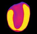
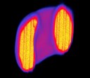
R4 microPET, a nuclear medicine modality, is capable of visualizing and detecting (static and dynamic) functional and physiological responses of a living organism non-invasively such as detecting cancer growth and metastasis, and inflammation due to cerebral and cardiac infarcts. Another area where PET modality plays a critical role in is its usefulness for drug discovery and screening due to advanced developments of kinetic modeling which are crucial for visualizing and quantifying specific drug efficacy. Finally, another aspect of R4 microPET is the translational capability. Translational aspect of small animal PET provides impetus for as well as expedites discovery of life saving drugs by quickly moving drug testing from preclinical animal models to human clinical trials.


