Research
Our research in pictures
The dynamic nucleolus of cultured rat cortical neurons. |
|
||||
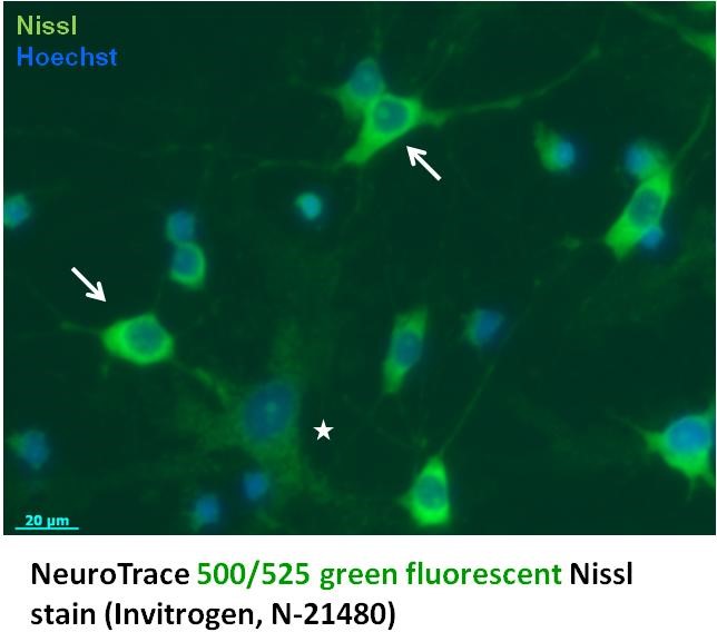
Ribosome staining in primary rat hippocampal cultures reveals high ribosome content in neurons (arrows) and relatively lower ribosome content in astrocytes (*). |
|
||||
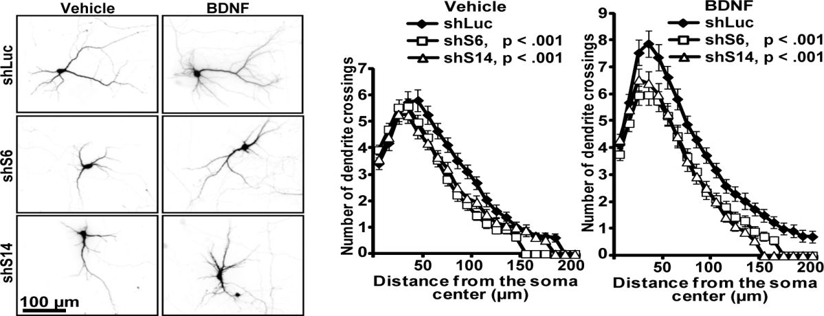
Anti-dendritic effects of knocking down ribosomal proteins S6 or S14 in primary hippocampal neurons. |
Slomnicki et al., JBC, 2016 (https://www.ncbi.nlm.nih.gov/pubmed/26757818) |
||||
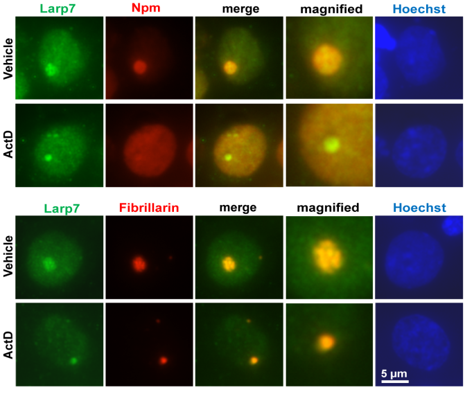
LARP7 resides in neuronal nucleoli. Note overlap of signal for LARP7 and the nucleolar FC marker, fibrillarin. Partial overlap with the GC marker NPM1 is also visible. LARP7 and fibrillarin appear to be co-localized even after inhibition of rRNA transcription using Actinomycin D. |
Slomnicki, Malinowska et al., MCP, 2016 (https://www.ncbi.nlm.nih.gov/pubmed/27053602) |
||||
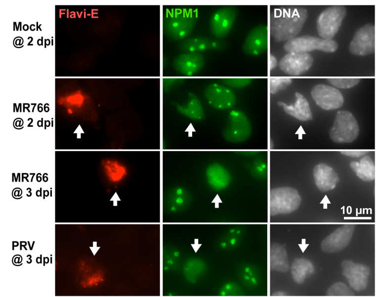
Zika virus infection disrupts nucleoli of embryonic cortical neurons. Infection of cultured cortical neurons from rat embryos with Zika virus (the African strain MR766 and the South American isolate PRVABC59 of the Asian Zika strain induced nucleoplasmic translocation of the nucleolar marker NMP1. |
Slomnicki et al., Sci. Rep., 2017 (https://www.ncbi.nlm.nih.gov/pubmed/29192272) |
||||
|
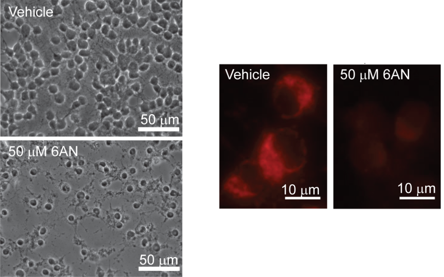
Inhibition of the pentose phosphate pathway using the NADPH antimetabolite 6-amino-NADP (6AN) induces cell death and mitochondrial damage in cultured rat oligodendrocyte precursor cells. Phase contrast images are after 24 h treatment with 6AN. Drop in mitochondrial potential (as indicted by red signal of the TMRM H+ probe) is observed after 6 h treatment with 6AN.
|
Kilanczyk et al., ASN Neuro, 2016 (https://www.ncbi.nlm.nih.gov/pubmed/27449129) |
||||

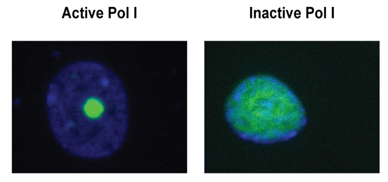 Upon 1 hr treatement with Actinomycin D to block Pol1 (0.05 mg/ml), the nucleoli of cultured rat cortical neurons disintegrate as demonstrated by a nucleoplasmic transclocation of the immunofluorescence for the nucleolar marker NPM1.