BIO-Imaging
Molecular Imaging
Molecular imaging methods that visualize the structure and function of the living body are widely used in clinical and biomedical research settings. Imaging techniques such as computed tomography (CT), plain film radiography, magnetic resonance (MR), and nuclear medicine provide in vivo data for measuring real-time responses in disease progression and therapeutic discovery studies. Nuclear medicine and molecular imaging have long been recognized to be of great value in cancer research and could be particularly beneficial for expediting the availability of translational results in response to an infectious outbreak. Research within the CPM is working to leverage these powerful approaches by developing predictive animal models of infectious diseases using molecular imaging modalities that—until now—have not been available to study pathogens requiring advanced biocontainment.
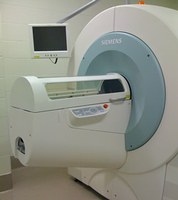
PET/SPECT imaging provides a new opportunity to study, in real-time, disease pathophysiology associated with pathogen replication and dissemination along with host response to infection. Our BIO-Imaging Core houses a Siemens Tri-modality scanner allowing for microPET, microSPECT, and microCT on multiple animal models for research utilizing BSL2, BSL3, and Select Agent pathogens.
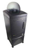
Our BIO-Imaging Core also houses a Caliper Life IVIS Spectrum capable of live-animal fluorescence and bioluminescent imaging providing another powerful approach for the study of pathogen and host cellular responses in real-time.
Microscopy
Zeiss epifluorescence and confocal microscopes are available for cellular imaging within BSL-3 containment. Each instrument is configured for live imaging with multiple plate formats.
Siemens Trimodal (PET/CT/SPECT)
- A unified control of PET, SPECT, and CT data acquisition
- LSO PET detector technology for high resolution (10%)
- New PET and SPECT acquisition and processing technology for improved count rate performance
- Unique PET transmission method for faster and more accurate attenuation correction and CT-based attenuation correction for PET imaging
- Advanced multi-pinhole SPECT collimators for improved sensitivity and spatial resolution
- Novel CT automated zoom control for optimized field of view and resolution, and variable-focus x-ray source with a maximum resolution of 15 microns
IVIS Spectrum
- Multi-modality instrument with optical technologies: high-sensitivity in vivo imaging of fluorescence and bioluminescence
- High throughput (5 mice) with 23 cm field of view high resolution (to 20 microns) with 3.9 cm field of view
- Twenty eight, high efficiency filters spanning 430 – 850 nm
- Support spectral unmixing applications
- Ideal for distinguishing multiple bioluminescent and fluorescent reporters
- Optical switch in the fluorescence illumination path allows reflection-mode or transmission-mode illumination
- 3D diffuse tomographic reconstruction for both fluorescence and bioluminescence
- Ability to import and automatically co-register CT or MRI images yielding a functional and anatomical context for enhanced scientific discovery.
Microscopy
Our Zeiss AxioObserver and Confocal microscopes are available to research clients with “Direct Use” or full “Fee-4-Service” options. Discounts are available for long-term, time-lapsed experiments.
Molecular Imaging
The IVIS Spectrum is available with “Direct Use” or “Fee-4-Service” options for research clients. The Siemens Trimodal is available on a full-service, daily-rate basis.
Please contact us for additional information regarding use, scheduling, and current pricing.
William E. Severson, Ph.D.
Director, Shared Resources
Center for Predictive Medicine
Phone: (502) 852-1546
Email: William Severson
Jon Gabbard, Ph.D.
Program Manager
Center for Predictive Medicine
Phone: (502) 852-1365
Email: Jon Gabbard
Bitplane, Imaris 3D&4D Image Processing & Analyses Software
Center for Molecular Imaging Innovation and Translation, About Nuclear Medicine and Molecular Imaging
Caliper, In Vivo University, Imaging Resources
Molecular Expressions, Optical Microscopy Primer
Molecular Expressions, Live-Cell Imaging
Zeiss, On-Line Campus
Molecular Imaging
Imaging techniques such as computed tomography (CT), plain film radiography, magnetic resonance (MR), and nuclear medicine provide in vivo data for measuring real-time responses in disease progression and therapeutic discovery studies. Nuclear medicine and molecular imaging have long been recognized to be of great value in cancer research and could be particularly beneficial for expediting the availability of translational results in response to an infectious outbreak in accordance with the FDA regulatory requirements for pre-clinical trials. Research within the Center are leveraging these powerful approaches by developing predictive animal models of infectious diseases using molecular imaging modalities that—until now—have not been available to study pathogens requiring advanced biocontainment.
To effectively use molecule imaging in the study of an infectious agent in basic and preclinical studies, not only must the animal model be a representative host for emulating the course of disease in humans, but also the imaging modality should have well-understood parameters and benchmarks for analyzing the data to draw meaningful conclusions. Additionally, small animal imaging requires:
- Establishment of normal anatomy by CT
- Distribution and uptake by various organs of the radiotracer
- Efficient methods of image post-processing and analysis
- Consequence of radiotracer uptake period on signal-to-noise ratio
- Comparison of intraperitoneal and intravenous injection of radiotracer
- Influence of respiratory gating on image quality
The following videos focus on using the Zeiss microscope in CTRB 6th Floor, Room 635


