Laser-Induced Fluorescence/Dispersed Fluorescence Spectroscopy
Reactive chemical intermediates, especially free radicals, are of crucial importance to combustion and atmospheric chemistry. In our High-Resolution Spectroscopy Lab, we use laser-induced fluorescence/dispersed fluorescence (LIF/DF) technique to study the structure and dynamics of free radicals. They are usually produced under jet-cooled conditions using laser photolysis, discharge, pyrolysis, or laser ablation followed by free-radical reactions.
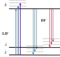
In the LIF experiment, molecules or free radicals are excited from the ground state to a high-energy state by laser and total fluorescence emission from the high-energy state to lower states are collected and detected. By scanning the frequency/wavelength of the laser, energy level structure of the high-energy state can be mapped out. In the DF experiment, the frequency of the excitation laser is parked on a LIF transition, while the fluorescence is dispersed and detected. The red-shift of the fluorescence signal with respect to the pump frequency gives the lower-state energy structure. The intensities of the LIF and the DF transitions provide further information about the studied molecule, such as the geometric structure of the molecule and the symmetries of the upper and lower states.

Figure 1. A principle of LIF/DF spectroscopy.
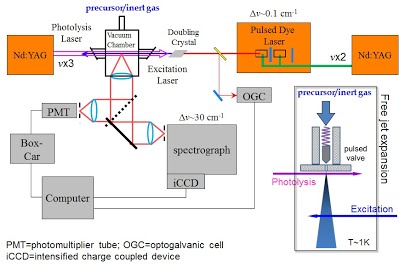
In our experiment, free radicals are generated in a supersonic jet expansion. The seeded flow of precursor molecules was expanded through a pinhole valve into the vacuum chamber to reach low temperature (~1 K). A Nd: YAG laser is used for photolysis of the precursors (using its 355 nm third harmonic or 266 nm fourth harmonic) or ablation (using its 1064 nm fundamental) followed by free-radical radiations. A dye laser pumped by the second harmonic of a Nd: YAG laser was used to excite the LIF transitions. The laser-induced fluorescence was collected by a lens system perpendicular to both the excitation laser beam and the jet expansion and detected by a photomultiplier tube (PMT). In the DF experiment, the fluorescence is focused into and dispersed by a monochromator, and detected by an intensified charge-coupled device (CCD) camera.

Figure 2: Experimental setup of LIF/DF spectroscopy.
The inset shows the supersonic expansion nozzle and the arrangement of photolysis and excitation laser beams.
