Tissue Ischemia and Energy Metabolism Laboratory
In Focus: Tissue Ischemia, organ preservation, wound care
Sufan Chien, MD
Professor
Division of General Surgery
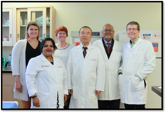
Our group has four scientists (Sufan Chien, MD, Harshini Sarojini, PhD, Girish Kotwal, PhD, and Mohammad Bayat, PhD) and two administrat assistants (Cheyrl Busch, BS and Lorrain Chian, MA) (Fig. 1). We have a special interest and dedication to tissue ischemia because this is the final pathway for all traumas and diseases that kills a patient. Despite more than a century of research and clinical efforts, no effective technique has been developed to combat ischemic damage except general supportive measures such as blood transfusion and oxygen therapy. The first approach was via organ preservation then expanded to other ischemic conditions. We have used various techniques and each has resulted in significant or unique contribution. The following is a brief description of several projects.
1. Extension of organ preservation time for transplantation
Organ transplantation is one of the most successful medical stories of the 20th century, but transplantation is also a victim of its own success, with demand for organs far exceeding supply. Extensive research in the past half century has failed to extend heart and lung preservation time even by 1 hour, no matter what technique or solution is used. The introduction of extended donor criteria now means that the limitations of cold storage have probably been reached. Our group focused on live-like autoperfusion because this was the most natural way of preservation. The multiple organ preservation developed by our group removed the heart, lungs, liver, pancreas, duodenum, and kidneys while the heart was still beating and lungs still oxygenating. Although the technique was delicate (Dr. Shumway called it “as complex as an emergency patient”), it kept the heart and lungs for more than 24 hours with good tissue structure and function—a result no other technique can match even nowadays (Fig. 2). Due to the technical difficulty and enormous human labor involved, it had a hard time to obtain continuous funding. Now, the issue of donor organ scarcity has become more critical, normothermic machine perfusion of individual organs or even the whole body has seen a rebound, and these procedures are far more expensive than multiple organ autoperfusion preservation. This approach may be worth further exploration again when new funding is available.
2. Enhancing energy production with fructose diphosphate
Although multiple factors are involved in organ damage during preservation, diminution of cellular ATP content is the most critical factor resulting in final irreversible organ damage. This is especially true for the heart: While other abdominal organs such as the liver and kidney can maintain membrane function and cell structure in the absence of substantial ATP reserve, depletion of ATP reserves in the heart results in ischemic contracture, because the actin-myosin cross bridge cannot be dissociated. Thus, even a small extension of preservation time will be highly significant. Because during the first step in glycolysis, glucose is converted into fructose 1,6-diphosphate (FDP), which consumes 2 ATP molecules. Using FDP directly can bypass the ATP-consuming steps thus increasing glycolytic energy production. Despite the numerous studies related to this chemical, a great deal of controversy exists. In particular, its ability to cross the cell membrane is under constant debate. Our effort was to investigate the potential usage of FDP in hypothermic heart preservation, and to study the mechanisms by which FDP enhances organ survival. We studied the effect of FDP on heart preservation and examined the passive permeability property of FDP. These results extended our understanding of FDP in organ preservation, which is still continuing nowadays.
3. Development of intracellular energy delivery
The intracellular energy delivery is the most recent but by far the most difficult task we have ever tackled. Due to the central role of ATP in cell metabolism and survival, tremendous efforts have been carried out to provide ATP to combat tissue ischemia. A direct intravenous infusion of ATP would be a simple solution. However, it is well known that strongly charged molecules like ATP normally cannot pass the membrane bilayer in sufficient quantities to satisfy tissue metabolic requirements. Furthermore, the half-life of free ATP in blood circulation is very short, limiting its efficacy as a bioenergetic substrate. After tremendous efforts, we have developed a technique for intracellular delivery of Mg-ATP by using specially formulated, highly fusogenic, unilamellar lipid vesicles. Because these vesicles have a composition similar to the cell membrane, they fuse with the cell membrane when they get into contact with them and deliver the content into the cytosol. The results have been very encouraging: In vitro studies indicated a rapid fusion with the endothelial cells, protection of endothelial cells, and cardiomyocytes during ischemia. In vivo studies have shown promising results in hemorrhagic shock, isolated organ preservation, and dramatically enhanced full-thickness skin wound healing in various animal models—granulation tissue starts to appear within 24 hours. It keeps growing and covers the wound cavity within a few days (Fig. 3). Reepithelialization tunneled through the granulation tissue. The top of the granulation tissue finally falls off, revealing perfectly healed wound. This is mainly achieved by massive macrophage trafficking, in situ proliferation, and direct collagen production—a process never seen or reported in the past. This technique has been funded by the NIH for many years and it has the potential to reduce or eliminate many detrimental effects caused by ischemia/hypoxia and our group is vigorously pursuing it in various applications.
4. Our new expansion of wound care research
In recent years, we have become more aggressive in pursuing wound care by incorporating two new directions: low-level laser therapy (LLLT) and newer animal models for better mechanistic study of chronic wounds and for more critical wound dressing tests. This is because human chronic wounds are highly diversified with various pathophysiology and treatment requirements. We have added LLLT to enhance our intracellular ATP delivery treatment hoping to obtain better results. At the same time, we have developed a newer technique to dramatically extend the neuroischemic time in our minimally invasive rabbit ischemic ear model. The new model is aimed to provide a new approach for mechanistic study and to give new wound dressing more critical tests.
Research supports:
Active
1. A new technique for diabetic foot ulcers (NIH).
Pending:
1. A long lasting neuroischemia in diabetes (NIH).
2. Rapid tissue regeneration for battlefield trauma )DOD).
Recently completed
1. Towards a universal chronic wound model (NIH).