Sample Studies and References
MicroPET Imaging
I. The Use of Dynamic Positron Emission Tomography Imaging for Evaluating the Carcinogenic Progression of Intestinal Metaplasia to Esophageal Adenocarcinoma
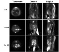
Study: Micro-PET images of rat after esophagoduodenal anastomosis (EDA).Sham: sham operation. W: week. M: month. The distribution of 18F-FDG in esophageal tissue progressively increased along the time course of the EDA model, 1 week esophagitis, 1 month esophagitis. Arrow: uptake of 18F-FDG by esophageal tissue after EDA.
Division of Surgical Oncology, Division of Diagnostic Radiology, Division of Gastroenterology/Hepatology, and Anatomic Pathology, University of Louisville School of Medicine
Reference : Yan Li, et al., Cancer Investigation, 26:278–285, 2008 Apr-May http://www.ncbi.nlm.nih.gov/pubmed/18317969
II. Positron emission tomography with fluorodeoxyglucose-F18 in an animal model of mania
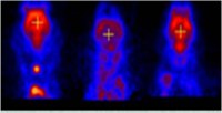
A B C
Coronal brain images in false color. A) Control animals receiving regular food and ICV artificial cerebrospinal fluid (aCSF) B) ICV ouabain treated animals C) Animals receiving lithium in their food and ICV ouabain
Study: Utilizing [18F-FDG] PET to visualize glucose uptake in animals receiving intracerebroventricular (ICV) ouabain. Lithium treatment did not alter 18FDG uptake, ICV ouabain treatment was associated with a significant reduction in 18FDG uptake compared with control and lithium treatment. Lithium pretreatment prevented the ouabain effect.
Department of Psychiatry and Behavioral Sciences, Department of Psychology, Diagnostic Radiology, Department of Obstetrics and Gynecology, University of Louisville School of Medicine, Louisville,
Reference : Rif S. El-Mallakha, et, al., Psychiatry Res. 2008 Oct 18
MicroCT imaging
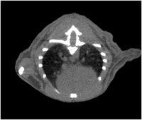
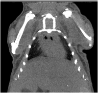
Reference link:
Optical imaging
I. IVIS imaging system:
IVIS Imaging technology provides the most sensitive imaging systems in the market - for both fluorescence and bioluminescence. IVIS molecular imaging systems are designed to enable researchers to identify disease pathways, determine mechanisms of action, evaluate efficacy of drug compounds, and monitor lead candidates' effects on disease progression in living animals - making in vitro to in vivo translational research a reality. Reference link: http://www.caliperls.com/products/optical-imaging/ivis-spectrum.htm
II. Maestro system:
CRi's patented imaging solutions reveal otherwise hidden signals and structures in biological samples. Detect, distinguish and quantitate signals with unsurpassed sensitivity and resolution. Make discoveries, accelerate drug development, and improve medical evaluation with superior imaging solutions that span histopathology, molecular biology, oncology and diagnostic imaging. Reference link: http://www.cri-inc.com/applications/in-vivo.asp