MicroCat II
|
|
|---|
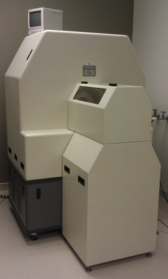
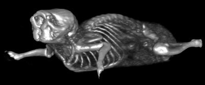
Siemens MicroCAT II is a dedicated and stand-alone CT scanner for small animals. With manual configurations between the detector and the X-ray source, the image resolution could vary from highest resolution of 27microns to the lowest resolution of 100 microns. With small dimensions inherent in small rodent subjects (20-30 g for mice and 200 250 g for rats), Siemens MicroCAT II has the capabilities and hardware to resolve and visualize small volume of rodent. MicroCAT II is an important piece of imaging modalities for preclinical research due to its dual functions that it plays in molecular imaging field. MicroCAT II allows researchers to acquire accurate attenuation maps for attenuation correction for both PET and SPECT. Another function that MicroCAT II plays is its role as a research tool for visualizing and quantifying anatomical structures of small animals non-invasively. With the principal output of CT images being the linear attenuation density functions (Hounsfield Unit), it is a superb modality for visualizing and quantifying bone density and integrity, and air-to-soft-tissue ratios for lung imaging.
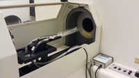
Images are acquired via a rotating X-ray detector and sources as they spin about a horizontal bed (or a stationary target). With respect to this particular MicroCAT II scanner, standard tungsten 40W-anode X-ray source (maximum operating energy at 80kVp and 500 micro-amperes) and a small pixels CCD detector (highest resolution at 27 micron [512 x 774] and lowest resolution at 100 micron [2048 x 3096]) are the principal data acquisition devices. The X-ray source provides high photon flux with large cone-beam angles, thereby improving spatial resolution during high magnification scans. Respiratory inlets and outlets for anesthesia could be easily inserted into the scanner via the small opening under the handle of the protective lead canopy. The scanner has build-in reconstruction algorithms (performed by COBRA reconstruction software) and capabilities to perform gated CT imaging (respiration only). The reconstructed images could be easily transferred and visualized via Siemens software.


