Fluorescence Imaging
 |  |
|---|
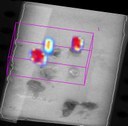
VisEn FMT 2500 LX (Fluorescence Molecular Tomography) is a dedicated in vivo fluorescence tomographic imaging system for rodents, which allows investigators to acquire calibrated quantitative 3D images. The FMT 2500 LX is operating under a four near-infared (NIR) -channels format, in which it is capable to excite fluorophores emitting 660-, 700-, 775-, and 805-nm wavelengths (excitation lights at 635, 670, 746, and 790 nm, respectively). By having multiple excitation channels, this construction improves the flexibility of the system. Moreover, with the build-in advanced 3D image reconstruction algorithms, the FMT 2500 LX allows investigators to accurately record and quantify in vivo tomographic fluorescence site-specific light emission from a target region. Because of the tomographic imaging capability, this unique feature also allows easy multi-modality fusion studies.
The principal sources of excitation lights are produced by four separate laser diodes (emitting photon radiation at 635-, 670-, 746-, and 790-nm wavelengths). During a data acquisition phase, a subject will be administered with a fluorescence-dyed labeled compound and a near-infrared (NIR) transilluminated laser diode is automatically positioned from the ventral side of the subject and the excited fluorescence output could then be collected at the dorsal side of the subject via a thermoelectrically cool CCD camera. Multiple calibrated projections of the excited fluorescence output from the subject could be recorded and the tomographic images could then be reconstructed from the multi-projected images. The output tomographic images could be exported or saved in a standardized medical file format called DICOM (Digital Imaging and Communications in Medicine) for post-processing. The FMT 2500 LX also has a built-in subject-holding cassette compartment and pressurized isoflurane inlet and outlets for easy subject loading and maintenance for imaging.
FMT modality is a unique optical imaging device which offers principal investigators with unprecedented opportunities to acquire 3-dimensional image data. Three-dimensional dataset allows pinpointing an exact location of the light emission sources, such as a tumor (similar to PET and SPECT) and notwithstanding this feature also allows researchers to make exact quantitative analysis on their target tissues. For example, using Prosense750 and MPSense680, FMT 2500 LX was used to image disease progression of rheumatoid arthritis in mice. The modality effectively discriminated and isolated regional recovery during the study's therapeutic phase. The 3D-mode could pinpoint the amount (or volume) of recovery.